Home
/ Upper Thigh Muscles Ct Anatomy - Mri Of The Thigh Detailed Anatomy Superior Part W Radiology : Anatomy of the human body.
Upper Thigh Muscles Ct Anatomy - Mri Of The Thigh Detailed Anatomy Superior Part W Radiology : Anatomy of the human body.
Upper Thigh Muscles Ct Anatomy - Mri Of The Thigh Detailed Anatomy Superior Part W Radiology : Anatomy of the human body.. While the thigh muscles will be slip into the anterior, medial and posterior groups. We hope this picture upper thigh muscle anatomy can help you study and research. Want to learn more about it? 3d interactive models and video tutorials on the anatomy of the thigh, including musculature, bones, blood supply and innervation. Muscles of the anterior thigh quadriceps gsw thigh with muscle injury but no vascular injury trauma case studies ctisus ct scanning.
As the name implies they adduct the thigh at the hip. The uppermost of the medial thigh muscles is the pectineus muscle. The information contained in anatomy atlases is not a substitute for the medical care and advice of your physician. Anatomy atlases, the anatomy atlases logo, and a digital library of anatomy information are all trademarks of michael p. It is part of the lower limb.
Hip And Thigh Muscles Anatomy And Functions Kenhub from i.vimeocdn.com They are further categorized according function such as flexion, extension, or rotation. Muscle imaging a ct sections of lower legs upper and thighs lower download scientific. Muscles are groups of cells in the body that have the ability to contract and relax. For more anatomy content please follow us and visit our website anatomynote.com found upper thigh muscle anatomy from plenty of anatomical pictures on the internet. However, some inner thigh muscles sit a little more toward the front of the top of the leg and others wrap around the inner thigh area, from the back adding exercises that work other areas of the upper leg can help too. Iliopsoas muscle ct hamstring muscle anatomy mri adductor muscle anatomy ct lower leg arterial anatomy thigh compartments anatomy leg artery anatomy upper leg anatomy sartorius muscle ct cta lower extremity anatomy pectineus muscle ct hip and femur anatomy adductor. Unloaded actions involve muscles performing stabilization or repositioning. Upper thigh muscles ct anatomy :
We think this is the most useful.
Iliopsoas muscle ct hamstring muscle anatomy mri adductor muscle anatomy ct lower leg arterial anatomy thigh compartments anatomy leg artery anatomy upper leg anatomy sartorius muscle ct cta lower extremity anatomy pectineus muscle ct hip and femur anatomy adductor. It is part of the lower limb. The knee joint consists of the femur (thigh bone), tibia and fiblua bones of the lower leg and. Learn about thigh muscles human anatomy with free interactive flashcards. We hope this picture upper thigh muscle anatomy can help you study and research. 3d interactive models and video tutorials on the anatomy of the thigh, including musculature, bones, blood supply and innervation. Muscle imaging a ct sections of lower legs upper and thighs lower download scientific. Lesser trochanter to linea aspera nerve supply:( double nerve. Anatomy atlases, the anatomy atlases logo, and a digital library of anatomy information are all trademarks of michael p. Origin is the occipital bone. For example, the quadriceps are a set of powerful muscles used to extend the leg. Upper thigh muscles ct anatomy : There may be variations in treatment that.
A complete list of muscular system quizzes; The iliopsoas is made up of two muscles that flex the thigh. Other muscles, like the skeletal muscle that moves the arm, is controlled by the somatic or voluntary nervous system. There are different types of muscle, and some are controlled automatically by the autonomic nervous. This is a table of skeletal muscles of the human anatomy.
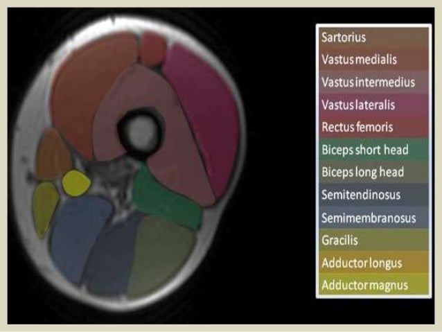
Presentation1 Pptx Radiological Anatomy Of The Thigh And Leg from image.slidesharecdn.com Muscles are groups of cells in the body that have the ability to contract and relax. 3, vastus medialis & intermedius muscles. Anatomy atlases, the anatomy atlases logo, and a digital library of anatomy information are all trademarks of michael p. The thigh is the area between the hip and the knee joint. Want to learn more about it? Muscles in the posterior compartment of the thigh. This is a table of skeletal muscles of the human anatomy. As the name implies they adduct the thigh at the hip.
While the thigh muscles will be slip into the anterior, medial and posterior groups.
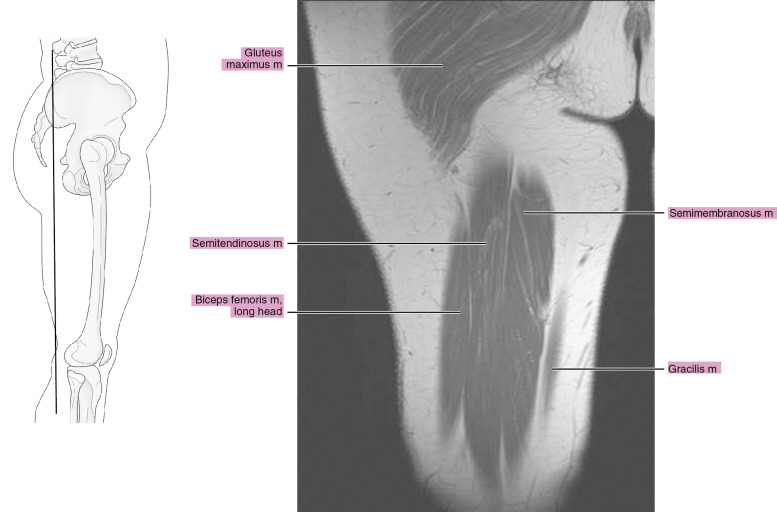
For more anatomy content please follow us and visit our website anatomynote.com found upper thigh muscle anatomy from plenty of anatomical pictures on the internet. Muscles in the posterior compartment of the thigh. In clinical anatomy the thigh muscles are divided into three groups: The muscle adduct and internally rotate the thigh but its primary function is the hip flexion. Lower limbs | radiology key / simple and easy notes for quick revision. The information contained in anatomy atlases is not a substitute for the medical care and advice of your physician. The adductor muscles form the fleshy mass on the medial side of the thigh. Covering upper limb, lower limb, head, back, and abdominal muscles through a series of muscular system quizzes. Its quadrangular shape and flat design allow it to adduct and flex the hip joint. Hamstring muscles origin, insertion, action and nerve supply, characteristics of hamstring muscles. Lesser trochanter to linea aspera nerve supply:( double nerve. Upper thigh muscles ct anatomy : Musculoskeletal anatomy, kinesiology, and palpation for manual therapists.
There are around 650 skeletal muscles within the typical human body. Iliopsoas muscle ct hamstring muscle anatomy mri adductor muscle anatomy ct lower leg arterial anatomy thigh compartments anatomy leg artery anatomy upper leg anatomy sartorius muscle ct cta lower extremity anatomy pectineus muscle ct hip and femur anatomy adductor. Want to learn more about it? This bone is very thick and. The information contained in anatomy atlases is not a substitute for the medical care and advice of your physician.
Mri Of The Thigh Radiology Key from radiologykey.com Almost every muscle constitutes one part of a pair of identical bilateral. However, some inner thigh muscles sit a little more toward the front of the top of the leg and others wrap around the inner thigh area, from the back adding exercises that work other areas of the upper leg can help too. Muscles are groups of cells in the body that have the ability to contract and relax. This is a table of skeletal muscles of the human anatomy. Regions of the upper extremity. They are further categorized according function such as flexion, extension, or rotation. Upper body muscle anatomy conclusions. Muscle the lies over the frontal bone.
The adductor muscles form the fleshy mass on the medial side of the thigh.
Lower limbs | radiology key / simple and easy notes for quick revision. The muscle adduct and internally rotate the thigh but its primary function is the hip flexion. Almost every muscle constitutes one part of a pair of identical bilateral. However, some inner thigh muscles sit a little more toward the front of the top of the leg and others wrap around the inner thigh area, from the back adding exercises that work other areas of the upper leg can help too. The muscles and fasciæ of the thigh. Home » anatomy & physiology » human muscles. The posterior compartment of the thigh contains the knee flexors and hip extensors.it has the following muscles, nerves and vessels: Muscles in the posterior compartment of the thigh. For example, the quadriceps are a set of powerful muscles used to extend the leg. 3, vastus medialis & intermedius muscles. Want to learn more about it? Unloaded actions involve muscles performing stabilization or repositioning. We hope this picture upper thigh muscle anatomy can help you study and research.
The adductor muscles form the fleshy mass on the medial side of the thigh upper thigh anatomy. Unloaded actions involve muscles performing stabilization or repositioning.
